Radiology Tech Interview Questions: What Employers Test
Radiology tech interview questions assess both technical expertise and patient care skills. Expect questions about radiation safety protocols, imaging equipment operation, patient positioning techniques, and emergency response procedures. Employers want technologists who understand ALARA principles, can operate multiple imaging modalities, communicate effectively with anxious patients, and troubleshoot equipment issues while maintaining diagnostic image quality and strict safety standards.
This guide covers essential competencies including radiation protection, imaging techniques across X-ray/CT/MRI modalities, patient interaction strategies, and quality control procedures. Build your foundation with medical imaging career resources.
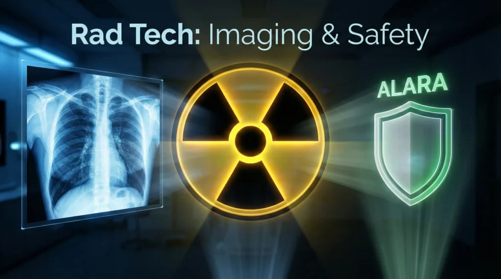
Understanding ALARA and Radiation Protection
What does ALARA mean and how do you implement it daily?
ALARA stands for As Low As Reasonably Achievable – a practical mindset for minimizing radiation exposure to patients, staff, and yourself while still producing diagnostic images. In day-to-day work, it comes down to three habits: time (use the shortest appropriate exposure and avoid repeats by positioning carefully the first time), distance (maximize separation from the source whenever you can), and shielding (use appropriate protective barriers and patient shielding when indicated and permitted by your facility’s policy).
Patient-specific measures include collimating tightly to region of interest limiting scatter radiation, selecting appropriate technical factors (kVp/mAs) for patient size avoiding unnecessary dose, and documenting pregnant status asking female patients of childbearing age before abdominal imaging. Personal protection requires wearing dosimeter badge monitoring occupational exposure, positioning behind protective barrier during exposures, and never holding patients during imaging using immobilization devices instead. Quality assurance ensures equipment calibration maintaining optimal performance with minimum dose and regular phantom testing verifying image quality at established exposure levels.
How do you protect pediatric patients from radiation?
Children require extra protection due to higher radiosensitivity and longer life expectancy for potential effects. Technical adjustments include reducing technical factors significantly – pediatric protocols typically use reduced technique factors compared with adult settings based on age/size guidance, increasing kVp when possible providing better penetration with lower dose, and using automatic exposure control (AEC) with pediatric settings preventing overexposure. Shielding emphasizes gonadal protection using shaped lead shields for reproductive organs whenever they’re within or near primary beam, thyroid shielding for chest and cervical spine imaging, and breast shields for female adolescents during thoracic procedures.
Image-gently campaign principles guide practice: scan only when necessary ensuring imaging truly changes management after consulting radiologist, scan only indicated region avoiding “just in case” additional views, and scan with child-sized settings using established pediatric protocols not adult techniques. Parent involvement includes allowing parent to stay with child when safe providing comfort reducing need for repeat images, explaining procedure to both child and parent reducing anxiety and movement, and positioning assistance from parent rather than technologist holding when appropriate minimizing staff exposure while supporting child.
Describe your radiation safety monitoring procedures.
Personal monitoring requires wearing dosimeter badge on collar outside lead apron measuring effective dose to most radiosensitive organs, checking badge monthly reviewing exposure reports and investigating any unusual readings, and maintaining ALARA goals and keeping occupational exposure well below regulatory limits. Equipment monitoring includes daily warm-up procedures checking tube output and detector calibration, weekly phantom testing verifying image quality and automatic exposure control accuracy, and monthly quality control documenting results per ACR or state requirements.
⚠️ Dosimeter compliance: Badge must be worn during all fluoroscopy and portable procedures. Never share badges or wear outside radiation areas. Report lost or damaged badges immediately to radiation safety officer.
Area monitoring uses survey meter checking scatter radiation levels in control booth and adjacent areas, posting radiation warning signs at procedure room entrances, and maintaining protective barriers ensuring lead equivalency meets specifications. Documentation maintains exposure logs tracking technique factors and patient dose for CT studies, incident reports documenting equipment malfunctions or unusual exposures, and quality assurance records demonstrating compliance with regulatory standards during inspections by state health department or Joint Commission surveyors.
Imaging Modality Expertise
General Radiography and Fluoroscopy
Q: Walk through positioning a patient for a chest X-ray.
Systematic positioning ensures diagnostic quality. Patient preparation includes removing all metal objects and clothing above waist replacing with gown opening to back, verifying correct patient and exam using two identifiers, and explaining procedure including breath-hold instruction. PA (posterior-anterior) upright positioning places patient facing image receptor with chin elevated clearing lung apices, shoulders rolled forward moving scapulae off lung fields, and hands on hips with elbows forward preventing rotation.
Centering aligns the central ray perpendicular to T7 (around the inferior angle of the scapula), positions the top of the image receptor slightly above the shoulders to include the apices, and ensures patient centered to receptor preventing cutoff. Technical factors typically use a long SID to minimize magnification, a high kVp technique to penetrate the mediastinum, and exposure on full inspiration to demonstrate adequate lung expansion. Quality check verifies symmetry (no rotation), adequate inspiration and penetration (vertebrae faintly visible through the heart silhouette), and proper collimation with lung fields fully included and no extraneous anatomy.
Computed Tomography Scanning
Q: How do you prepare a patient for CT with contrast?
Pre-scan assessment is critical for safety. Screen for prior contrast reactions and relevant allergies, and confirm kidney function and other risk factors per facility protocol. Review medications that may require special handling around contrast administration (for example, certain diabetes medications) and follow your site’s guidance. Verify fasting status if your department requires it for the specific study.
IV access establishes an IV that is appropriate for power injection (often in an antecubital vein), tests patency flushing with saline ensuring no infiltration, and secures catheter preventing dislodgement during table movement. Patient education explains warming sensation during injection, metallic taste possibility, and importance of remaining still during scan. Post-procedure monitoring includes observing for any immediate or delayed reactions per protocol, encouraging hydration when appropriate, and providing clear instructions on when to seek medical attention (for example, rash, swelling, or breathing difficulty).
Q: Explain CT dose reduction techniques.
Dose optimization balances image quality with radiation safety. Protocol selection uses appropriate scan range limiting coverage to diagnostic area not scanning beyond clinical indication, selects tube current modulation adjusting mAs automatically based on patient attenuation reducing dose in thinner body regions, and chooses iterative reconstruction reducing noise allowing lower mAs while maintaining image quality. Technical parameter optimization lowers tube current (mAs) for pediatric and thin patients using size-based protocols, adjusts pitch increasing pitch above 1.0 for screening exams when high resolution not critical, and limits multiphase scanning avoiding unnecessary pre-contrast or delayed phases.
Size-specific dose estimate (SSDE) accounts for patient size calculating actual dose received by small or large patients more accurately than standard CTDI, displays dose metrics recording CTDIvol and DLP for every exam documenting radiation exposure, and tracks cumulative dose monitoring patients receiving multiple CT exams identifying high-dose patients. Quality initiatives include establishing diagnostic reference levels comparing facility doses to national benchmarks, regular protocol review optimizing techniques as scanner technology improves, and technologist education training on dose reduction features ensuring proper utilization maintaining ALARA compliance.
MRI Safety and Screening
Q: What MRI safety screening questions are critical?
Metallic implants pose greatest risk in powerful magnetic field. Pacemaker/defibrillator screening determines if device MRI-conditional (safe at specific field strengths with restrictions) or contraindicated – MRI-conditional devices may be safe only under specific conditions and typically require clearance and monitoring per protocol. Aneurysm clips ask about brain surgery as ferromagnetic clips can move causing hemorrhage while titanium clips generally safe. Cochlear implants verify MRI compatibility as older models contain ferromagnetic components though newer implants often MRI-conditional.
Metallic foreign bodies include orbital metal fragments screening welders and metal workers with orbit X-rays before scanning, shrapnel or bullet fragments determining location and composition, and surgical hardware like joint replacements (usually safe titanium) versus older stainless steel implants. Pregnancy discusses risk-benefit requiring radiologist approval for first trimester scans, avoiding gadolinium contrast unless essential due to unknown fetal effects. Claustrophobia assesses anxiety level offering open MRI when available, using mirror allowing patient to see out of bore, and considering anxiolytic medication for severe cases with physician order.
Q: How do you handle a patient who becomes claustrophobic during MRI?
Prevention strategies reduce anxiety before scanning. Pre-scan preparation includes facility tour showing scanner and explaining procedure, positioning discussions describing how patient will lie and how long scan takes, and comfort measures offering blankets, pillows, and music selection. Communication system demonstrates call button patient can squeeze if becoming anxious, establishes checking-in protocol technologist talking to patient between sequences, and family presence allows loved one in scanner room when safe providing reassurance.
During-scan interventions if patient becomes anxious involve immediate response stopping scan and talking to patient through intercom, gradual desensitization pulling patient partially out allowing them to see room and calm down, and position adjustment trying feet-first rather than head-first entry when anatomy permits. Medication options include an anxiolytic given pre-scan if ordered by a clinician (with appropriate post-visit safety planning), conscious sedation monitoring vitals throughout procedure with RN present, and postponement rescheduling for another day if patient cannot complete exam. Alternative imaging considers if different modality (CT, ultrasound) could answer clinical question avoiding MRI entirely when patient unable to tolerate despite interventions.
Patient Care and Communication Skills
Q: Describe managing an uncooperative pediatric patient.
Child-friendly approach builds cooperation. Age-appropriate communication uses simple terms explaining “picture machine” for young children, shows equipment letting child touch or explore when safe, and engages imagination suggesting they pretend to be statue or superhero staying still. Distraction techniques include videos or music playing child’s favorite show during procedure, toys or books keeping child occupied during positioning, and parent participation having parent talk to child or hold hand when possible.
Immobilization devices when necessary include Pigg-O-Stat for chest X-rays securing infant upright safely, Velcro straps for extremities gentle restraint preventing movement, and positioning sponges supporting body parts comfortably. Timing considerations schedule early morning when child well-rested and cooperative, work quickly minimizing time child must hold still, and take breaks if procedure lengthy allowing child to relax between images. Never forcefully restrain child risking injury and traumatizing patient – consult radiologist about sedation if truly uncooperative and exam medically necessary ensuring safe completion while preserving child’s trust in healthcare.
Q: How do you explain procedures to patients with limited English?
Language barriers require creative communication. Interpreter services use hospital language line calling professional interpreter for complex consent or instructions, schedule in-person interpreter for lengthy procedures like fluoroscopy when real-time communication essential, and avoid using family members especially children as interpreters due to accuracy and privacy concerns. Visual communication demonstrates procedure using gestures and pointing, shows sample images helping patient understand what you’re looking for, and uses translation apps for simple phrases though verify accuracy with back-translation.
Written materials provide procedure instructions in patient’s language printing commonly-needed languages in advance, use picture-based handouts illustrating positioning and breathing instructions, and give discharge papers in native language ensuring post-procedure care understood. Universal techniques include speaking slowly and clearly avoiding medical jargon, using simple yes/no questions patient can answer with head nod, and confirming understanding asking patient to demonstrate position or repeat instructions ensuring safety despite language differences showing cultural competence and patient-centered care.
Equipment and Quality Assurance
What do you do if equipment malfunctions during a procedure?
Systematic response ensures patient safety and minimal disruption. Immediate actions include stopping procedure prioritizing patient safety over completing exam, removing patient from equipment if any safety concern exists, and assessing situation determining if minor fixable issue or major malfunction requiring service. Communication notifies supervisor immediately about equipment down for scheduling adjustments, informs radiologist if critical exam requiring alternative solution, and updates patient explaining delay and offering alternative appointment or different modality when appropriate.
Troubleshooting basic issues attempts simple solutions like restarting system which often resolves software glitches, checking connections ensuring cables properly seated and power supply functioning, and consulting user manual reviewing error codes and recommended actions. Service call documents problem clearly describing error messages and what happened before malfunction, takes equipment out of service posting signage preventing use until repaired, and schedules service prioritizing based on equipment availability and exam backlog. Never attempt major repairs beyond scope of practice – specialized biomedical engineers should handle complex issues maintaining warranty and ensuring safety.
Describe your quality control responsibilities.
Daily QC ensures consistent performance. Morning warm-up procedures include running equipment through warm-up cycle before first patient allowing tube and detector to stabilize, checking image quality using test phantom verifying no artifacts or calibration drift, and reviewing prior day’s pending exams ensuring no technical repeats from previous shift. Documentation maintains QC log recording test results and any corrective actions, tracks repeat rate analyzing why images repeated identifying training needs, and monitors equipment utilization noting any recurring problems requiring service attention.
Periodic testing includes weekly checks like automatic exposure control verification using phantom ensuring consistent exposures, monthly evaluations of collimation accuracy confirming light field matches radiation field, and quarterly inspections by medical physicist measuring radiation output and image quality parameters. Participate in quality improvement reviewing departmental quality metrics with team, suggesting protocol optimizations based on clinical feedback, and staying current on ACR standards attending annual competency updates maintaining technical excellence throughout career demonstrating commitment to professional development and patient safety.
Radiology Knowledge Assessment
20 Quick Review Questions
1. ALARA stands for?
- As Low As Reasonably Achievable
- Always Limit All Radiation Amounts
- Automatic Low-dose Adjustment Required Always
- Advanced Level Appropriate Radiation Assessment
2. Three principles of radiation protection are?
- Time, distance, shielding
- Speed, accuracy, safety
- Quality, quantity, compliance
- Exposure, positioning, documentation
3. For a standard PA chest radiograph, technologists typically use?
- A short SID because it’s faster
- A long SID to reduce magnification
- Any distance; it doesn’t affect image geometry
- The shortest distance to reduce dose
4. Gonadal shielding used when?
- Gonads within or near primary beam
- All imaging procedures
- Never used anymore
- Only for pediatric patients
5. MRI contraindication includes?
- Dental fillings
- Non-MRI-conditional pacemaker
- Titanium joint replacement
- Previous CT scan
6. CT contrast requires checking?
- Renal function (creatinine/eGFR)
- Blood pressure only
- Patient weight only
- No screening needed
7. Dosimeter badge worn?
- Inside lead apron
- Outside lead apron at collar level
- On wrist
- Only during fluoroscopy
8. Pediatric imaging requires?
- Same technique as adults
- Reduced technique factors using pediatric protocols
- Higher radiation dose
- No special considerations
9. Image Gently campaign focuses on?
- Pediatric radiation dose reduction
- Adult imaging quality
- MRI safety
- Equipment maintenance
10. Collimation purpose is?
- Limit radiation to area of interest, reduce scatter
- Increase image brightness
- Speed up procedure
- Protect equipment
11. ARRT certification requires?
- Passing the exam and completing required continuing education
- One-time exam only
- No continuing education
- Medical degree
12. Pregnant patients for abdominal X-ray?
- Always refuse imaging
- Consult radiologist, use alternative if possible
- Proceed normally
- Only after delivery
13. Automatic exposure control (AEC)?
- Terminates exposure when sufficient radiation detected
- Manually set by technologist
- Not used anymore
- Only for CT
14. PA chest should show?
- 5 posterior ribs
- Adequate inspiration (assessed by visible posterior ribs)
- 15 ribs minimum
- Any number acceptable
15. In CT, using a higher pitch generally means?
- The table moves faster relative to the beam, often lowering dose
- Radiation dose always increases
- It indicates a scanner error condition
- Image quality always improves
16. Claustrophobic MRI patient may benefit from?
- Forcing completion
- Pre-medication, open MRI, feet-first positioning
- Refusing to scan
- Sedation always required
17. Language barrier addressed by?
- Asking family to interpret
- Professional interpreter services, visual aids
- Proceeding without explanation
- Refusing to scan
18. Equipment malfunction requires?
- Continuing to use
- Stop procedure, notify supervisor, take out of service
- Patient fixing it
- Ignoring issue
19. Daily QC includes?
- Warm-up, phantom testing, reviewing repeat rate
- No testing needed
- Only when problems noticed
- Annual check sufficient
20. Repeat images should be?
- Routine for all exams
- Minimized through proper technique, tracked for QA
- Never acceptable
- Not documented
Common Radiology Tech Interview Questions
🎓 Should I bring my ARRT certification to the interview?
Yes, bring current ARRT card and any specialty certifications (CT, MRI, mammography) demonstrating credentials. Many employers verify certification status, so having documentation ready shows professionalism. If recently certified and card not yet arrived, bring temporary verification from ARRT website showing active status and exam pass date.
🧲 How do I demonstrate experience with multiple modalities?
Discuss specific procedures performed in each modality quantifying volume like “performed 20-30 chest X-rays daily” or “cross-trained in CT completing 100+ exams during rotation.” Mention equipment manufacturers and models showing familiarity with different systems. If limited in certain modalities, emphasize quick learning ability and willingness to obtain additional certifications showing growth mindset.
⚠️ What if asked about radiation exposure I caused?
Frame honestly as learning experience using STAR method. Describe situation (what happened), task (your responsibility), action (how you addressed it – notified radiologist, documented incident, reviewed protocol), and result (what you learned and changes made to prevent recurrence). Shows accountability, commitment to safety, and professional maturity. Never hide mistakes – demonstrates integrity valued in healthcare.
🛡️ How technical should answers be for MRI safety?
Balance clinical application with technical understanding. Mention specific contraindications (non-MRI-conditional pacemakers, certain aneurysm clips, orbital metal fragments) showing knowledge, but focus on screening process and patient safety protocols demonstrating practical skills. If interviewing for MRI-specific position, expect deeper technical questions about field strength, SAR limits, and quench procedures requiring specialized MRI knowledge.
💬 Should I discuss difficult patient scenarios?
Absolutely – prepare 2-3 specific examples showing patient care skills. Good scenarios include managing anxious patient (demonstrated communication reducing fear), adapting for disability (modified positioning for wheelchair-bound patient), or handling emergency (patient became unstable during procedure showing quick thinking). Emphasize patient-centered approach, teamwork with nurses/radiologists, and maintaining composure under pressure proving you handle challenging situations professionally.
Landing Your Radiology Tech Position
Success with radiologic technologist interview questions requires demonstrating comprehensive radiation safety knowledge, technical proficiency across imaging modalities, excellent patient communication skills, problem-solving ability with equipment issues, and commitment to quality assurance. Employers value technologists who prioritize ALARA principles, adapt techniques for diverse patient populations, work collaboratively within healthcare teams, and maintain current certifications through continuing education.
Prepare thoroughly by reviewing anatomy and positioning, practicing STAR responses for behavioral questions, organizing examples of patient interactions and technical challenges, and researching facility’s imaging equipment and patient demographics. Bring your ARRT certification, maintain professional demeanor, and ask informed questions about department protocols and growth opportunities. For additional strategies, visit radiology career advancement guides demonstrating your dedication to diagnostic imaging excellence and patient-centered care.
⚠️ Disclaimer: The interview strategies, sample answers, and negotiation tips provided in this guide are for educational purposes only. Hiring decisions are subjective and vary by company and industry. While these strategies are based on professional HR standards, they do not guarantee a specific job offer or result.